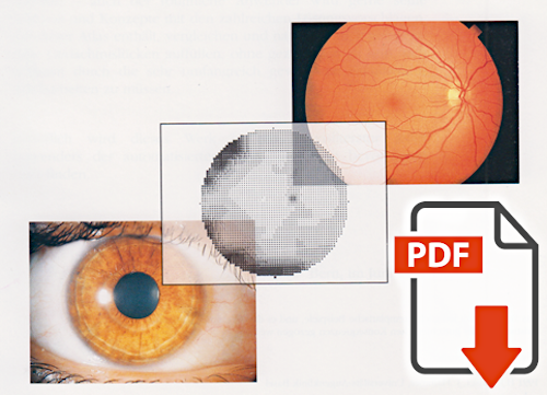
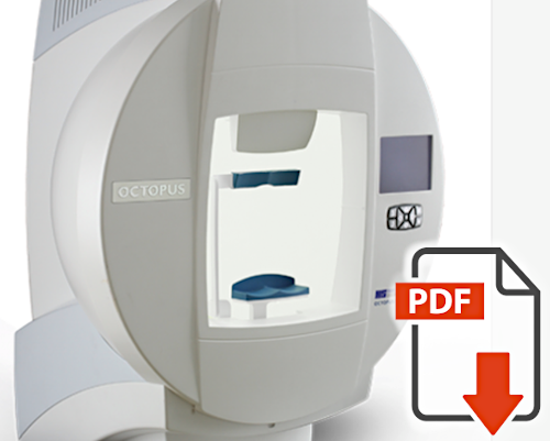
Automated perimetry
 J Flammer: The concept of visual field indices |
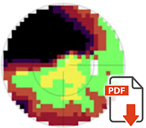
The result of a static perimetry consists of many numbers. An automatic perimeter can represent this graphically, e.g. in the form of a gray scale. However, to facilitate a quantitative comparison of visual fields, Josef Flammer developed and introduced the so-called visual field indices. You can find his corresponding original description here. |
 J Flammer, F Jenni, H Bebie, B Keller: The Octopus Glaucoma G1 Program |
The first automatic perimeter (Octopus) was developed by Prof. Franz Fankhauser. However, the measurements took a long time at that time. Equidistant measurement grids were used, which is not optimal for glaucoma. When J. Flammer joined Fankhauser's research group, he devoted himself specifically to glaucoma perimetry. This work resulted in, among others, the octopus glaucoma program G1 and G2. Today, decades later, this program is still in use worldwide. Very often today it is also used for perimetry of patients with other diseases. Here you can find the original description of the glaucoma program G1. |
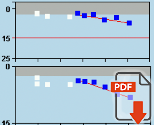
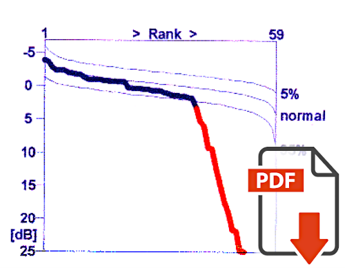 H Bebie, J Flammer, Th Bebie: The cumulative defect curve: separation of local and diffuse components of visual field damage |
In the past it was assumed that glaucoma caused only local deficits in the visual field, i.e. only scotomas. Diffuse deficits were attributed entirely to other causes, e.g. cataract. Today, we know that glaucoma-related nerve fiber defects, and thus visual field defects, can be both local and quite diffuse. Quantifying such diffuse defects and separating them from the local defects is not straightforward. Therefore, Bebie et al. introduced the cumulative defect curve (so-called Bebie curve). This allows to visually separate the two types of defects. |
 HJ Kaiser, J Flammer: Visual Field Atlas |
This visual field atlas by Kaiser et al. helps to interpret the visual fields and relate them to diseases. It is intended for ophthalmologists, neurologists, optometrists, perimetrists and interested patients. Visual field examples of more than 40 different diseases are shown. This atlas has been published in English, German and Spanish. HJ Kaiser and J Flammer: Gesichtsfeldatlas / Visual Field Atlas / Atlas de Campo Visual. Buser Printing Company, Basel Switzerland, 1991 |
 A Weijland, F Fankhauser, H Bebie, J Flammer: Automated perimetry, Visual Field Digest |
For some of us, an automated perimeter is a "black box". This book "Automated Perimetry, Visual Field Digest" explains the technical and physiological basics. It explains why the perimeter measures what and how. This allows the user to properly understand the results. It is written by authors who are pioneers in perimetry research and the development of automated perimetry. |
 J Flammer, M Drance, L Augustiny, A Funkhouser: Quantification of Glaucomatous Visual Field Defects with Automated Perimetry |
In this original paper, Flammer et al. describe the different ways to visualize and quantitatively describe a visual field. This includes numerical grids, gray levels, three-dimensional sensitivity mountains, visual field indices, and cumulative relative frequencies. Most of the methods developed then and described here have found their way into clinical routine. |
 PA Rabineau, J Flammer: Sources of Errors in Automated Static Perimetry |
When interpreting visual field defects, the question often arises whether and to what extent a defect is not or not only from glaucoma, but possibly completely or partially from another disease. Equally important, however, is the question of whether the deficits are real or possibly just artifacts. In this paper, Rabineau et al. describe the various possible sources of error in perimetry, such as incorrect refraction, dirty contact lenses, anatomical obstructions, or cooperation problems, etc. |
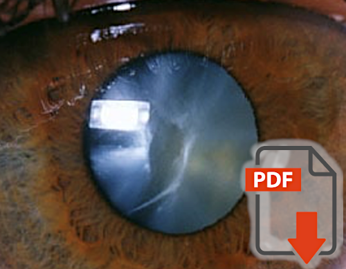 K Yao, J Flammer: Relationship cataract density and visual field damage |
Glaucoma patients often have a more or less advanced cataract. This of course has an influence on the visual field. Yao et al. have shown that the influence of a cataract on the visual field is quite diffuse, i.e., it does not mimic scotomas. Cataract-induced glaucoma damage correlates with the severity of lens opacification. After cataract surgery, the visual field improves but does not completely normalize. The reason is that artificial lenses are optimized more for visual acuity but less for the (peripheral) visual field. |