| When quantification of ocular blood flow was still difficult, Flammer and coworkers concluded ocular blood flow from the relationship between peripheral blood flow and visual field behavior in glaucoma. Meanwhile, there have been major advances in the methodology for measuring various parameters of ocular blood flow. Thus, it is now possible to inversely deduce blood flow in other organs from blood flow in the eye. In particular, dynamic vascular analysis of the retina gives information about the state of vascular endothelial cells throughout the body. This helps various specialists, such as nephrologists, neurologists or the otologists. The connections between the eye and the heart are particularly well studied. |

E Waldmann, P Gasser, B. Dubler, Ch Huber, J Flammer:
Silent myocardial ischemia in glaucoma and cataract patients |
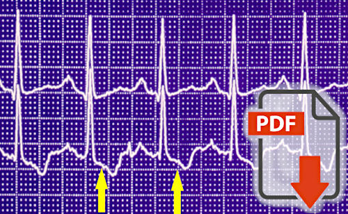
Waldmann and co-authors used long-term ECG to show that glaucoma patients go through periods of silent (painless) ischemia more frequently than healthy control subjects. Within glaucoma patients, this was again significantly more common in patients with normal tension glaucoma than in patients with high tension glaucoma. This demonstrates the existence of a systemic vascular component in glaucoma, particularly normal tension glaucoma. The observation that these episodes of myocardial hypoxia in glaucoma rarely occur during exercise but more frequently at rest and during sleep indicates that it is rarely caused by atherosclerosis but more frequently by vascular dysregulation. |

J Flammer, K Konieczka, RM Bruno, A Virdis, AJ Flammer, S Taddei:
The eye and the heart |
Ophthalmologists Flammer and Koniecka and cardiologists from Taddei's group describe and illustrate the connections between the eye and the heart in this review. The blood vessels in the retina are suitable for vascular diagnostics because they are easily accessible optically and are not innervated autonomously. This allows, among other things, a global functional analysis of the vascular endothelial cells. Morphology, diameter, and behavior of retinal blood vessels also provide cardiologists with very important information about the health status of blood vessels in general. Dilated retinal veins have a particularly strong prognostic significance. This dilatation, in turn, is often due to increased retinal venous pressure. |